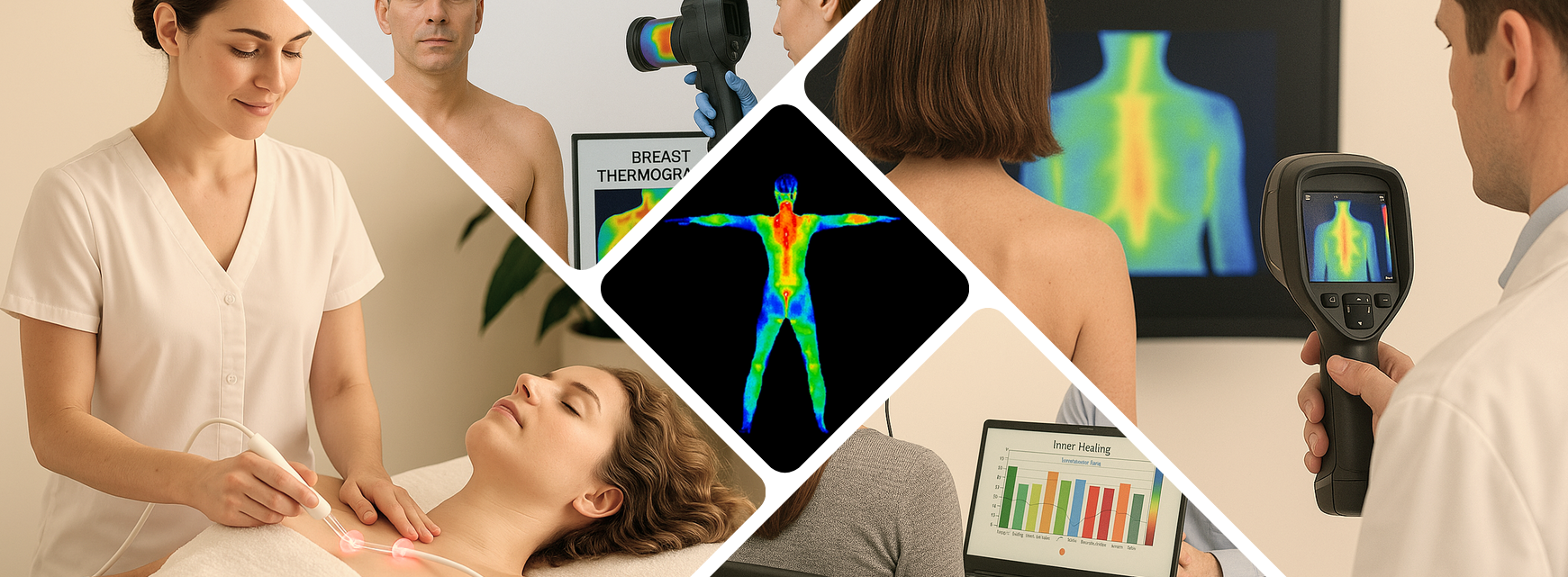
Breast Thermography is a 15 Minute Non Invasive Test of Physiology.
Breast thermography is a 15-minute, non-invasive screening test designed to remove much of the uncertainty that often surrounds breast disease detection. Instead of relying solely on structural imaging—like mammography, ultrasound, or MRI—thermography focuses on physiology. Unlike traditional exams that rely only on structure, thermography monitors physiology — changes in temperature, vascular development, and heat patterns — giving you and your doctor a powerful early-warning system. It interprets how breast tissue behaves, not just how it looks. This distinction is powerful, because physiological changes such as inflammation, vascular development, tissue irritation, and abnormal heat patterns frequently appear far earlier than visible structural changes.
Thermography uses medical-grade digital infrared cameras to measure subtle temperature variations across the surface of the breasts. These temperature patterns are influenced by underlying tissue health and blood flow. When tissue becomes stressed, inflamed, hormonally stimulated, or begins forming abnormal vascular networks, the breast often produces distinguishable heat signatures. These signatures may indicate physiologic changes long before anything becomes large enough to appear on a traditional structural scan.
One of the greatest strengths of breast thermography is its ability to establish a personalized baseline. Your first scan creates a unique thermal “map” of your breasts—similar to a fingerprint. During subsequent scans, this baseline becomes the comparison point for detecting even the smallest changes in temperature, symmetry, or vascular activity. Because the body tends to maintain consistent thermal patterns over time, a shift from your established norm can be a valuable early signal worth further evaluation. This is why thermography is strongly recommended on an annual basis, with a three-month follow-up after the initial scan to confirm stability.
The test itself is fast, gentle, and completely comfortable. There is no radiation exposure, no compression, no contact with equipment, and no invasive procedures. You simply stand in a temperature-controlled room while a series of infrared images are taken from several angles. The process is private, quiet, and designed to keep you at ease throughout the brief imaging session.
Once the images are captured, they are analyzed by trained thermographers who specialize in interpreting thermal physiology. They look for indicators such as asymmetric heating, increased vascular patterns, areas of persistent inflammation, or localized cool regions known as “thermal cold spots” that can correspond to underlying dysfunction. Every scan is archived securely so your breast-health history can be monitored year after year.
Thermography is not a replacement for mammography or other imaging tools, and it is not intended to function as a stand-alone diagnostic method. Instead, it is a complementary screening option that enhances early detection efforts by focusing on physiologic activity rather than structure alone. Many women—especially those under 50, those with dense breast tissue, those seeking radiation-free screening options, and those committed to prevention-focused care—find that thermography provides a level of insight and peace of mind they simply cannot get from structural imaging by itself.
By combining thermography with traditional medical imaging and regular clinical exams, you and your provider gain a more complete picture of breast health. This layered approach supports proactive wellness, encourages early detection, and empowers you to participate actively in your long-term breast-care plan.
How It Works
When breast tissue undergoes abnormal activity—such as cancerous growths, fibrocystic changes, inflammatory processes, infections, hormonal stimulation, or increased blood flow—its thermal signature begins to shift in ways that can be detected long before structural changes appear. These physiologic shifts alter the way heat is distributed across the surface of the breast. Areas experiencing accelerated cell growth often produce excess heat, while certain types of abnormalities may create regions that appear cooler due to vascular compromise or tissue disruption. Because the body’s thermal patterns are remarkably consistent from one person to the next, even subtle variances can carry important clinical meaning when they deviate from what is expected or from your documented baseline.
Using ultra-sensitive digital infrared cameras, breast thermography captures these temperature variations through a series of high-resolution thermal images. Unlike standard imaging tools that are designed to identify masses or physical abnormalities, infrared thermography records patterns of heat that reflect underlying physiological activity. The technology is capable of detecting microvascular changes, inflammation, and subtle temperature asymmetries—factors that often precede structural changes by months or even years. This early physiologic awareness makes thermography an invaluable complement to traditional imaging and an empowering tool for women who want a more proactive and comprehensive approach to monitoring their breast health.
During your initial appointment, a detailed thermal map of your breast tissue is created. This first imaging session becomes your personal baseline—a unique thermal fingerprint that represents what is normal for your body at this moment in time. Unlike one-time structural scans, which only show a snapshot of anatomy, thermography’s strength lies in its ability to track functional changes over long periods. Once your baseline is established, it becomes the reference point for all future scans. This allows thermographers to monitor even the slightest shift in temperature patterns, vascular development, or regional asymmetry.
Because your baseline is so central to accurate interpretation, your second thermography session is typically scheduled about three months later. This follow-up scan is important, not because changes are expected, but to verify that your thermal patterns are stable and reproducible. If the two scans match—meaning no significant variations are present—you now have a reliable baseline that can be used for annual comparisons. If subtle changes appear, they are evaluated to determine whether they indicate normal physiologic fluctuation or whether further monitoring or clinical correlation is appropriate.
Year after year, repeated annual scans offer a powerful trend-tracking system. Instead of relying solely on structural imaging, which may only detect issues once they have progressed, thermography provides a dynamic and ongoing record of how your breast tissue behaves over time. This long-term physiologic monitoring is one of thermography’s greatest advantages, especially for women who want to detect subtle abnormalities early or who may have risk factors—such as dense breasts, family history, hormonal imbalance, chronic inflammation, or previous breast health concerns—that make traditional imaging less effective or more difficult to interpret.
What makes thermography especially meaningful is not simply the detection of heat, but the interpretation of thermal patterns. For example, irregular vascular development—such as new blood vessels feeding a specific region—can signal abnormal activity, because increasing blood supply is one way the body supports rapidly growing cells. Likewise, asymmetrical heating between the left and right breast may suggest hormonal influence or inflammation. Certain patterns of heat, cooling, or vascularity have long been studied and documented, allowing trained thermographers to recognize when a pattern appears benign, hormonal, suspicious, or clearly abnormal.
By capturing these changes consistently, thermography gives both patients and healthcare providers a more complete picture of breast health, allowing earlier intervention when necessary and peace of mind when patterns remain stable. For many women, especially those who want a radiation-free, contact-free, and non-invasive screening option, thermography becomes an essential part of their wellness strategy. It encourages awareness, accountability, and early action—a unique combination that empowers you to stay deeply connected to your own breast health in a way that no other single method can offer.

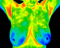
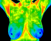
Normal
A normal breast thermography scan shows good thermal symmetry, meaning both breasts display balanced and harmonious temperature patterns without noticeable irregularities. There are no areas of excess heat, cooling, or abnormal vascular activity that would suggest inflammation, hormonal imbalance, or physiologic stress. The vascular patterns appear consistent, organized, and typical for healthy breast tissue, showing no signs of chaotic or newly formed blood vessels that might warrant further evaluation. Overall, a normal scan indicates stable physiology and provides a reliable baseline for future comparison, helping ensure that any subtle changes over time can be detected quickly and accurately.
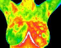
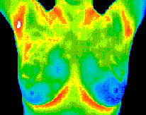
Fibrocystic Changes
Fibrocystic changes often appear on thermography as increased vascular activity, localized inflammation, or areas of heightened thermal response. In this case, the significant vascular patterning in the left breast warranted clinical correlation, which later confirmed that the findings were consistent with fibrocystic changes rather than a more concerning process. These benign but hormonally responsive changes can fluctuate over time, making regular thermographic monitoring especially useful. By scanning at appropriate intervals, a clear pattern can be established, allowing thermographers and clinicians to determine when the breast has reached a stable baseline suitable for reliable annual comparison and long-term breast-health tracking.
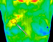
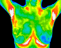
Early Stage Malignant Tumor
In early-stage malignant processes such as a small DCIS, thermography may reveal distinct physiological patterns that differ from benign changes. In this example, the imaging shows a clear vascular “feed,” indicating the development of new blood vessels supporting abnormal cellular growth. Surrounding this area is a discreet region of hypothermia, where the tumor disrupts normal heat distribution, creating a cooler focal point bordered by compensatory hyperthermia. These contrasting patterns—abnormal vascularity, localized cooling, and surrounding heat—form a recognizable thermal signature that warrants prompt clinical evaluation. Identifying such subtle changes early allows for timely follow-up imaging and medical intervention.
The Big Advantage
Thermography offers a powerful advantage in breast-health monitoring because it evaluates physiology rather than structure. While mammography looks for anatomical changes such as masses or calcifications, thermography focuses on how the breast tissue is functioning—specifically, whether there are patterns of heat, vascular development, or inflammation that differ from your normal baseline. This makes it an ideal complementary tool rather than a replacement. The two methods work together to give you and your healthcare provider a far more complete picture of what’s happening inside the breast.
Think of thermography as your first line of early detection. Physiologic changes often occur long before a tumor is large enough to be seen on a traditional mammogram. Increased blood flow, abnormal vascular feeding, temperature asymmetry, or inflammatory responses can create early warning signs that thermography is uniquely equipped to capture. By identifying these subtle changes early, thermography allows for proactive follow-up—whether that means additional clinical exams, ultrasound, mammography, or simply closer observation.
Research over several decades has shown that digital infrared imaging can detect abnormalities with accuracy comparable to mammography in some studies, and significantly better than clinical breast exams alone. When used alongside structural imaging, thermography strengthens the ability to identify issues earlier, track progression, and confirm when physiologic patterns remain stable over time.
Another major advantage is patient comfort. Thermography is completely non-invasive, radiation-free, contact-free, and painless. For women with dense breasts, implants, tenderness, or those seeking a radiation-free screening method, this makes thermography a valuable addition to their annual breast-health routine.
In short, thermography expands what traditional screening can reveal. By detecting physiologic change early and helping guide timely next steps, it empowers women to take a more active, informed role in protecting their long-term breast health.

Ideal For
-
Women under 50 whose breast density may limit mammography effectiveness.
-
Anyone seeking to monitor breast-health risk actively.
-
Those who prefer a completely radiation-free, contact-free procedure.
-
Patients invested in prevention, not just treatment.
What to Expect
We’ll take a series of infrared breast images, then compare them to your baseline and published normal patterns. If your scan shows abnormal signs — like asymmetric heating, unusual vascularity, or thermal cold spots — we’ll work with your physician for follow-up imaging, ultrasound, or other diagnostic tools. All scans are carefully archived so year-to-year tracking becomes a routine part of your breast-health strategy.
