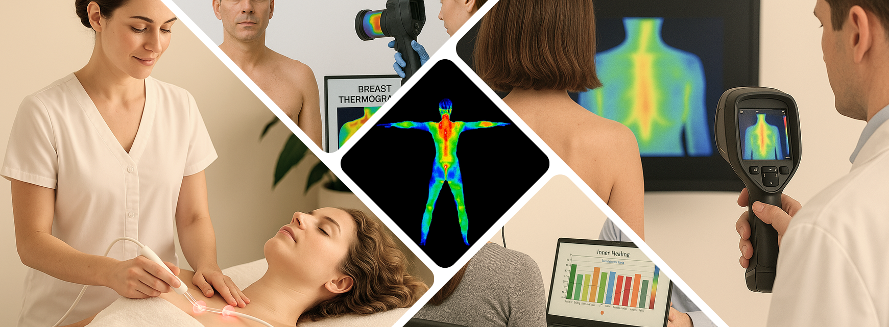
Understanding Deep Vein Thrombosis (DVT) and the Role of Infrared Thermography
Blood clots that form deep in the body—known medically as deep vein thrombosis (DVT)—are a serious health condition. They most often develop in the deep veins of the legs but can occur elsewhere. If a clot breaks free and travels to the lungs, it can cause a life-threatening pulmonary embolism (PE). Recognizing risk factors, spotting signs early, and using appropriate diagnostic tools are all critical.
In recent years, infrared thermal imaging has emerged as a valuable non-invasive adjunct in the diagnosis and screening of Deep Vein Thrombosis (DVT). While it is not a replacement for gold-standard vascular studies, it offers meaningful physiologic insight that can support early detection and clinical decision-making. DVT occurs when a blood clot forms in a deep vein—most commonly in the legs—restricting circulation and potentially leading to life-threatening complications such as pulmonary embolism. Because early symptoms can be subtle or mistaken for muscle strain, timely evaluation is critical.
Thermal imaging contributes to this process by capturing heat patterns associated with inflammation, impaired venous flow, and localized autonomic responses. Abnormal temperature increases or asymmetry between limbs may indicate underlying vascular changes consistent with DVT. Although these findings must be confirmed with Doppler ultrasound or additional diagnostic tests, thermography can help clinicians identify areas of concern sooner, prioritize urgent evaluations, and monitor physiological changes over time.
As part of a comprehensive assessment, thermal imaging adds an extra layer of visual information—enhancing awareness, supporting clinical judgment, and helping ensure that potential DVT cases receive prompt attention.
What is DVT and why is it dangerous?
When a blood clot forms in a deep vein—most commonly in the leg—it can significantly restrict normal blood flow. This reduced circulation often leads to local symptoms such as pain, swelling, warmth, and redness. While these issues are concerning on their own, the greatest danger comes from the possibility that the clot could break loose. If it travels through the bloodstream and becomes lodged in the lungs, it can cause a pulmonary embolism, a life-threatening medical emergency that requires immediate attention.
Understanding risk factors is essential for early recognition and prevention. Some of the most common contributors include prolonged immobility, such as long flights or extended bed rest; recent surgery or trauma; certain medical conditions that affect clotting; pregnancy; hormone therapies; smoking; obesity; and a personal or family history of clotting disorders. Athletes may also be at heightened risk during periods of intense training, dehydration, or injury.
Recognizing these risks enables clinicians and patients to stay alert to early warning signs and seek timely evaluation.
-
Surgical procedures, especially under general anesthesia and lasting more than about 20 minutes. Studies show that post-surgical patients — particularly older than 40 — may have DVT rates as high as 14–33% (after abdominal surgery) and nearly 50% (after hip surgery) if no prophylaxis is applied.
-
Immobility, obesity, recumbency or bed-rest.
-
Pre-existing conditions: cardiovascular disease, venous disorders, blood disorders (e.g., elevated fibrinogen), history of DVT/PE, cancer.
-
Inherited or acquired clotting-factor abnormalities (e.g., deficiencies in antithrombin III, Protein C, Protein S)
Because DVT can be "silent" (without obvious symptoms) yet have severe consequences, screening and early detection are vital.
Traditional diagnostic approaches
Historically, the gold standard for diagnosing Deep Vein Thrombosis (DVT) has been contrast venography, a procedure in which a contrast dye is injected into a vein and then imaged with X-ray to visualize blood flow. While venography provides highly accurate results and detailed visualization, it is also invasive, uncomfortable for the patient, and not without risk. The use of contrast dye may cause allergic reactions, kidney strain, or irritation at the injection site. Because of these limitations, venography—though reliable—is rarely the first choice in modern clinical practice.
More commonly, clinicians now rely on duplex ultrasonography, a non-invasive test that combines traditional ultrasound imaging with Doppler technology to assess both the structure of the veins and the movement of blood flow within them. Duplex ultrasound is safe, widely accepted, and highly effective in detecting clots in the major veins of the legs. However, it does have its constraints. Its accuracy may vary depending on the technician’s skill, the patient’s anatomy, or the location of the suspected clot—particularly in smaller distal veins or in individuals with significant swelling. Additionally, ultrasound may not always be immediately available in urgent or resource-limited settings.
Because both venography and ultrasound come with accessibility, cost, or practicality limitations, researchers and clinicians have increasingly explored alternative and complementary diagnostic strategies. Among those emerging tools is infrared thermography, which offers a non-invasive way to visualize physiological changes associated with DVT. Instead of examining structure directly, thermography evaluates temperature distribution and vascular patterns on the skin’s surface—changes that may reflect underlying inflammation, impaired venous return, or shifts in autonomic activity caused by a developing clot.
Although thermography is not intended to replace established diagnostic methods, its ease of use, lack of radiation, and ability to detect physiologic abnormalities early make it an appealing adjunct. In environments where rapid triage is needed—or where access to advanced imaging may be limited—it can serve as a valuable screening tool to help identify high-risk patients who require immediate follow-up testing.
As part of a comprehensive diagnostic approach, thermography adds another layer of insight, helping clinicians make faster, more informed decisions about which patients may need urgent ultrasound or additional vascular assessment.
What is Infrared (Thermal) Imaging and how might it help in DVT?
Infrared or thermal imaging works by detecting differential heat patterns across the surface of the body. Because the skin reflects underlying physiological activity—especially changes in blood flow, inflammation, and autonomic responses—thermography can reveal temperature variations that might otherwise go unnoticed. In the context of Deep Vein Thrombosis (DVT), the idea is straightforward: when a clot forms or deep-vein flow becomes impaired, the surrounding tissues often respond with inflammation or altered circulation. These changes can influence heat distribution, creating localized “hot spots” due to increased metabolic activity or, in some cases, cooler areas where circulation is reduced.
By capturing these subtle asymmetries between limbs or regions, thermal imaging may provide early clues that something is physiologically abnormal. While these findings are not diagnostic on their own, the presence of unusual heat patterns can prompt further evaluation, guide clinicians toward targeted testing, and help identify patients who may require immediate vascular assessment.
Some published studies have evaluated the use of liquid crystal thermography, thermo-camera profiles and combined approaches. A few noteworthy results:
-
In one study of 102 patients with suspected DVT, plethysmography had a sensitivity of 63% and specificity of 94%. A thermographic temperature-profile method showed 87% sensitivity but only 39% specificity; a thermo-camera method had 83% sensitivity and 55% specificity. When plethysmography + temperature-profiles + pattern recognition were combined, sensitivity reached 96% and specificity about 81%.
-
In another trial of 100 patients, liquid crystal thermography achieved a negative predictive value of 97% if done within one week of symptom onset. Duplex ultrasonography in that study had sensitivity ~93% and specificity ~91% for all thrombi.
The takeaway: thermal imaging shows promise as a screening tool—especially to rule out DVT when negative—but is not sufficiently specific to serve as a standalone diagnostic test when positive. A positive result still needs follow-up with duplex ultrasound or venography.
How to interpret thermal imaging results in this setting
-
A negative thermogram (i.e., no abnormal heat or cold patterns) within a week of symptom onset appears to strongly suggest the absence of DVT — reducing the need for more invasive tests in some patients.
-
A positive thermogram however, does not confirm DVT; it may prompt further evaluation. Because surface temperature changes can be caused by many other issues (infection, trauma, inflammation, superficial vein problems), specificity is limited.
-
Therefore: Thermography may serve as a first-line non-invasive screen, especially in settings where other diagnostic tools are not immediately available. But it should not replace standard investigations when suspicion is high.
If you or someone you know has risk factors for Deep Vein Thrombosis (DVT)—such as recent surgery, extended immobility, long travel, hospitalization, a prior history of clots, active cancer, hormone therapy, smoking, obesity, pregnancy, or an inherited clotting disorder—it’s important to take even mild symptoms seriously. DVT can develop quietly, and early signs are often subtle enough to be mistaken for a pulled muscle or general soreness. However, symptoms such as persistent leg swelling, new or worsening pain, warmth, redness, tenderness along a vein, or an unexplained difference between the two legs should never be ignored.
Seeking immediate evaluation is essential because early detection dramatically reduces the risk of complications. The greatest danger occurs when a clot breaks loose and travels to the lungs, causing a pulmonary embolism—a life-threatening emergency that may present with chest pain, shortness of breath, dizziness, or rapid heartbeat. Prompt assessment allows clinicians to perform appropriate testing, begin anticoagulation when needed, and prevent the clot from expanding or migrating.
Whether symptoms are mild or severe, acting quickly can make a major difference in outcome. When in doubt, it’s always safer to get checked. DVT is treatable—and early intervention is one of the most effective ways to protect your long-term health.
What this means for you
If you or someone you know has risk factors for DVT (recent surgery, immobility, previous history, cancer, clotting disorders, etc.), or is showing signs such as leg swelling, pain, warmth/redness, then immediate evaluation is important. Early detection can make a major difference.
If your clinic offers thermal imaging and you’re considering it for DVT screening, keep in mind:
-
Make sure patients understand that thermography is screening, not definitive diagnosis.
-
Use thermography mainly to stratify risk or exclude DVT in low-to-moderate suspicion cases.
-
Be prepared to order or refer for duplex ultrasound or venography if the thermogram is positive or if clinical suspicion remains high.
-
Ensure patients are properly informed about follow-up, risks of DVT/PE, and general preventive strategies (mobility, compression stockings, managing risk factors).
Preventive measures and best practices
Even before any screening or imaging, prevention is key. Here are some suggestions:
-
For surgical patients: use prophylaxis (mechanical and/or pharmacologic) as per guidelines, especially in longer procedures or high-risk individuals.
-
Encourage mobility and leg-movement exercises during periods of immobility (bed rest, long flights, sedentary jobs).
-
Maintain a healthy weight, manage cardiovascular risks (hypertension, high cholesterol, smoking), and treat underlying venous disease.
-
Stay vigilant for symptoms of DVT (leg swelling, pain or tenderness, warmth, discoloration) and PE (shortness of breath, chest pain, rapid heart rate). Seek immediate care if these arise.
Final thoughts
Deep vein thrombosis (DVT) is a serious and potentially life-threatening condition that deserves timely attention, careful evaluation, and proactive management. Although symptoms can begin subtly, the consequences of a missed or delayed diagnosis can be severe. A clot that forms in a deep vein may remain stable for a time, but it also carries the risk of breaking loose and traveling to the lungs, resulting in a pulmonary embolism. Because of this, clinicians must approach suspected DVT with vigilance, and patients must understand the importance of seeking prompt medical care.
In recent years, non-invasive screening tools such as infrared or thermographic imaging have emerged as helpful adjuncts in the evaluation process. These technologies offer a physiologic perspective rather than a structural one, capturing subtle temperature asymmetries and circulatory changes that may reflect underlying inflammation or venous impairment. In the right clinical context, thermography can provide valuable insight—helping clinicians identify patients who may be at low risk, guiding decisions about who needs further workup, and offering a visual aid that supports patient education and awareness.
However, it is equally important to recognize the limitations of thermographic screening. While it may help rule out DVT in select cases or assist with risk stratification, it does not replace established diagnostic methods such as duplex ultrasonography or, when necessary, contrast venography. When clinical suspicion is high, or when symptoms strongly suggest a clot, traditional imaging remains the gold standard. Thermography should be viewed as a complementary resource rather than a standalone diagnostic tool.
For clinicians considering the integration of thermography into their practice, thoughtful implementation is essential. Begin by educating patients about what thermography can—and cannot—detect. Clear communication builds trust and ensures that patients understand the purpose of the screening, how it fits into the broader diagnostic plan, and why follow-up testing may still be required. Use thermography selectively, focusing on cases where physiologic visualization can add actionable information, such as borderline symptoms, low-to-moderate clinical suspicion, or situations where repeated structural imaging is not feasible.
When abnormal findings appear on thermographic images, they should be interpreted as indicators that warrant further testing rather than definitive evidence of DVT. Following through with appropriate investigations—typically duplex ultrasound—helps confirm the diagnosis, guide treatment, and protect patient safety. When results are normal, thermography can provide reassurance and help clinicians determine whether additional evaluation is necessary.
Ultimately, early detection and prevention remain the strongest tools in reducing the complications associated with DVT. Encouraging patients to recognize their risk factors, understand common symptoms, stay active during long periods of immobility, and seek assessment when something feels “not quite right” can significantly reduce the incidence of severe outcomes. Coupling these strategies with thoughtful use of emerging technologies like thermography gives clinicians a more comprehensive, layered approach to vascular health.
As the landscape of diagnostic medicine continues to evolve, tools that enhance early recognition and improve patient engagement will continue to play an important role. Thermography, when used appropriately, can be one of those tools—supporting better decision-making, refining risk assessment, and contributing to safer, more informed care for individuals at risk of DVT.
Frequently Asked Questions
- Can a thermal imaging scan (thermography) reliably detect a deep vein thrombosis (DVT) in its early stage, and if so, what are the limitations?
- If I’ve had one DVT, what are the chances of having another clot in the future — and are there preventive measures beyond staying active and hydrated?
- How might external factors like long-haul flights, elevated altitude, or major surgery affect the risk of DVT — and what should a person do to mitigate that risk?
Q1. Can a thermal imaging scan (thermography) reliably detect a deep vein thrombosis (DVT) in its early stage, and if so, what are the limitations?
Answer:
Yes — thermography can detect temperature irregularities associated with increased blood flow or inflammation in the region of a developing clot. The article cites older research showing high sensitivity for thermographic screening of DVTs (for example a sensitivity of ~94 %) in some settings.
However, there are important caveats:
-
Thermography is non-specific — a hot spot might be inflammation from other causes, not necessarily a clot.
-
It cannot replace definitive imaging (such as duplex ultrasound or venography) when DVT is strongly suspected.
-
Its utility may depend on how early the clot has formed, the anatomical location (calf vs thigh vs pelvis), ambient temperature, patient factors (skin thickness, limb movement) and operator experience.
So the takeaway: thermography may be a useful screening adjunct, especially when traditional imaging is not immediately available, but it should not be relied on alone to rule in a DVT if clinical suspicion is high.
Q2. If I’ve had one DVT, what are the chances of having another clot in the future — and are there preventive measures beyond staying active and hydrated?
Answer:
Having a prior DVT does raise your risk of recurrence. For example, literature shows about 30 % of people may have a recurrent venous thromboembolism (which includes DVT or pulmonary embolism) in the ten years following an initial event. Beyond general lifestyle measures (keeping active, maintaining a healthy weight, avoiding prolonged immobility, staying hydrated) you might consider:
-
Discussing with your doctor whether you should have testing for inherited or acquired clotting disorders (especially if your first clot was unprovoked).
-
Using graduated compression stockings after an event, which may reduce risk of post-thrombotic syndrome and possibly recurrence.
-
Following up with your healthcare provider regarding the duration of anticoagulant therapy after your first clot (some cases may warrant extended treatment depending on risk factors).
-
Avoiding additional risk factors when possible (for example, smoking cessation, managing obesity, careful planning of long trips or hospital stays).
In short, yes — you do have a higher risk of a second clot, but by combining medical follow-up with smart preventive strategies you can reduce that risk significantly.
Q3. How might external factors like long-haul flights, elevated altitude, or major surgery affect the risk of DVT — and what should a person do to mitigate that risk
Answer:
External/temporary factors definitely play a role in DVT risk:
-
Long periods of immobility (such as during long flights or car trips) slow venous return, which increases the risk of clot formation.
-
Major surgery (especially orthopaedic, abdominal, or pelvic) is a known DVT precipitant because of vessel trauma, immobility, and changes in blood-flow and clotting post-surgery.
-
Elevated altitude and dehydration may further increase clot risk (less discussed in the blog but present in other sources).
What to do: -
For long flights/trips: get up and walk every hour if possible, if not then perform calf stretches and foot-circles; stay well hydrated; avoid alcohol; wear loose clothing.
-
If you’re going into major surgery, discuss prophylactic measures (such as anticoagulants, compression devices, early mobilization) with your physician.
-
For travel or when altitude is a factor: plan ahead — compression stockings may help; ensure frequent movement; ask your doctor if you have known clotting risk factors whether extra preventive measures are needed.
So external/in-motion factors do matter — and being proactive during these higher-risk situations is important.
