Digital Infrared Thermal Imaging in Sports Medicine & Musculoskeletal Care
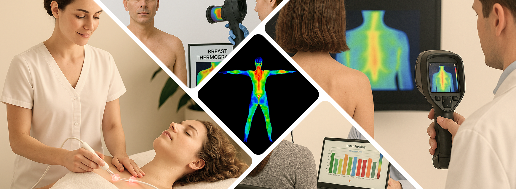
When it comes to diagnosing and managing sports injuries, clinicians are always seeking tools that are accurate, safe, and practical. Digital Infrared Thermal Imaging (DITI) checks every one of those boxes — and provides valuable insight into the body’s autonomic and sympathetic responses in ways no other test can. By visualizing surface temperature patterns, clinicians gain a clearer understanding of inflammation, circulation changes, nerve irritation, and other physiological responses that occur before structural damage becomes visible on traditional imaging.
In the world of sports medicine, where early detection and precise monitoring are essential, DITI has steadily earned its place as a trusted and highly effective resource. Athletic trainers, physical therapists, chiropractors, and sports physicians rely on thermographic imaging to help identify injuries earlier, document baselines, and monitor an athlete’s healing trajectory over time. Because it captures physiological changes rather than anatomical structure, DITI often highlights dysfunction before it progresses into a more serious or chronic condition.
Another major advantage is its versatility. Whether evaluating acute injuries like sprains, strains, and impact trauma, or chronic issues such as stress fractures, tendonitis, and nerve compression, DITI provides a visual map of thermal abnormalities that can guide further testing or targeted treatment. This makes it especially useful for shaping individualized recovery plans and for determining when an athlete is truly ready to return to play—helping reduce re-injury risk and improving long-term outcomes.
Because DITI is non-invasive, radiation-free, and highly portable, it integrates seamlessly into clinics, training rooms, and private practices. Athletes appreciate that the process is quick, comfortable, and requires no physical contact. For clinicians, the ability to capture reproducible, real-time physiological data supports better decision-making and elevates the standard of care.
DITI is not just an imaging option—it’s a powerful, forward-thinking tool for precision sports injury management.
Why DITI Matters in Sports Medicine
DITI isn’t just a diagnostic snapshot — it’s a dynamic tool that supports the entire lifespan of an athletic injury. From the very first sign of discomfort to the final clearance for competition, thermography provides clinicians with continuous, objective information they can rely on. Because it visualizes subtle physiological changes such as inflammation, vascular alterations, and autonomic responses, DITI reveals what the athlete’s body is doing beneath the surface long before structural imaging can.
This deeper level of insight allows providers to identify emerging problems early, track healing with measurable data, and make informed decisions about treatment intensity and progression. During rehabilitation, thermography helps confirm whether an athlete is responding appropriately to therapy or if adjustments are needed to prevent setbacks. When it comes time for return-to-play considerations, DITI adds another layer of confidence by highlighting whether the injured area has stabilized or if hidden dysfunction remains.
By offering real-time physiological feedback throughout recovery, DITI strengthens clinical judgment and enhances athlete safety at every stage.
DITI plays multiple roles throughout recovery, including:
-
Confirming a diagnosis by identifying abnormal thermal patterns that correlate with inflammation, nerve irritation, or vascular changes.
-
Identifying injury patterns early, even before structural changes appear on X-rays or MRIs.
-
Tracking healing over time, offering a visual timeline of progress that both athletes and clinicians can clearly understand.
-
Assessing treatment effectiveness, allowing providers to adjust therapies based on the body’s real-time response.
Conditions Commonly Evaluated With DITI
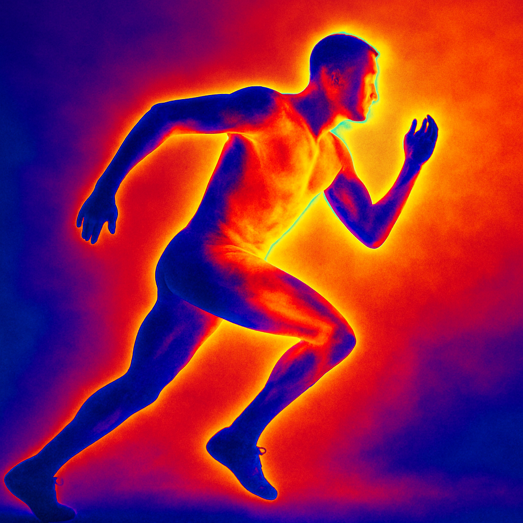
Sports medicine clinicians use DITI for a wide range of conditions, including:
-
Epicondylitis (tennis/golfer’s elbow)
One of DITI’s greatest contributions is its ability to detect post-traumatic pain syndromes such as Reflex Sympathetic Dystrophy (Complex Regional Pain Syndrome) and sympathetic-maintained pain — conditions that are both clinically complex and historically difficult to diagnose early. These pain syndromes involve abnormal, often exaggerated activity of the autonomic nervous system following an injury, surgery, or other trauma. Because the dysfunction is physiological rather than structural, traditional imaging tools like X-rays, CT scans, and MRIs generally fail to reveal the underlying problem. By the time structural changes appear — if they appear at all — the condition is often far more advanced.
Digital Infrared Thermal Imaging bridges this critical gap by offering a real-time, non-invasive view of the body’s autonomic responses. Instead of looking at bones or tissue, DITI evaluates temperature patterns and vascular reactions that reflect sympathetic activity and functional imbalances. Abnormal heat increases may indicate inflammation or autonomic hypersensitivity, while cooler regions often signify vasoconstriction or impaired blood flow. In post-traumatic pain syndromes, these distinctive thermal asymmetries frequently emerge early, sometimes before clinical symptoms escalate to severe pain or disability.
This makes DITI a powerful “first-alert” tool. Clinicians can detect subtle abnormalities at a stage when the condition is still highly treatable. By identifying these patterns early, providers can intervene quickly, potentially reducing the likelihood that the patient will develop chronic, long-lasting pain. This is particularly important in conditions like CRPS, where delayed diagnosis is strongly associated with poorer outcomes.
DITI essentially provides a window into the autonomic nervous system — something no other imaging modality does as safely, effectively, or consistently. With each scan, clinicians gain objective information about how the sympathetic nervous system is behaving on both a local and systemic level. For example, the imaging may reveal temperature differences between limbs, patchy vasomotor changes, or progressive heat shifts that correspond with worsening dysfunction. These patterns are not only diagnostic clues but also valuable indicators of severity and progression.
Beyond early detection, thermography is equally valuable throughout the treatment and recovery process. Because autonomic disorders fluctuate, a single snapshot is rarely enough to understand how the condition is evolving. DITI allows clinicians to track changes over time, documenting how the body responds to therapy, medication, nerve blocks, or physical rehabilitation. This serial imaging approach gives providers insight into whether symptoms are stabilizing, improving, or worsening — enabling more informed, timely adjustments to the treatment plan.
The ability to visualize objective physiological data also serves a crucial purpose for patients. Many individuals with CRPS or sympathetic-maintained pain struggle with validation because their symptoms can be severe even when traditional tests show “normal” results. Thermography bridges that gap by offering a clear, visual representation of their experience. This can strengthen physician–patient communication, support disability or insurance claims when necessary, and give the patient a greater sense of clarity and control.
In challenging, complex pain cases where uncertainty often slows diagnosis and treatment, DITI offers something rare: objective, reproducible, meaningful data. It doesn’t just complement clinical judgment — it enhances it, giving providers a clearer path toward early intervention, improved outcomes, and more effective long-term management.
How DITI Reflects the Sympathetic Nervous System
Thermal emissions from the skin are controlled by the sympathetic nervous system, which manages:
-
Dermal microcirculation
-
Vasomotor tone
-
Peripheral arteriolar constriction
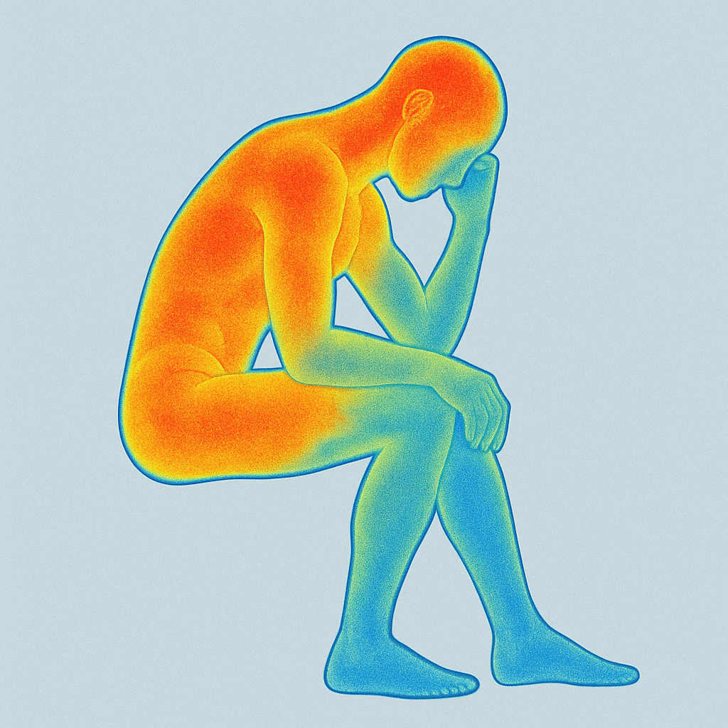
When sympathetic activity increases, vessels constrict, reducing heat at the skin’s surface (a hypothermic pattern). When sympathetic activity decreases — such as after injury or during receptor fatigue — vessels dilate and heat increases (a hyperthermic pattern).
These temperature changes create distinctive thermal maps that mirror the body's physiological responses to injury.
Understanding Pain Through Thermal Imaging
DITI does not image pain itself — instead, it visualizes the autonomic dysfunction associated with pain.
-Pain at the injury site often appears hotter (hyperthermic) because of decreased sympathetic function and the presence of biochemical substances like histamines and substance P.
-Referred pain often appears cooler (hypothermic) due to increased sympathetic activity.
-This makes DITI especially valuable for identifying myofascial pain, referred pain zones, and neurogenic dysfunction, which are not always visible on X-rays, MRIs, or ultrasounds.
Studies show strong correlation between thermal findings and patient-reported pain regions, supporting DITI as a dependable adjunct diagnostic tool.
DITI Is Backed by Leading Medical Organizations
DITI has been recognized for its diagnostic value for decades:
- 1987 – AMA Council on Scientific Affairs
- 1988 – Congress of Neurosurgeons
- 1990 – American Academy of Physical Medicine and Rehabilitation
- ACA Council on Diagnostic Imaging
Research consistently supports its reliability:
- 96% interobserver reliability in low back pain studies
- 98% test efficiency and 94% interrater reliability in knee pain studies
These findings demonstrate that thermography is not only clinically useful but also repeatable and objective — two qualities essential in sports injury evaluation.
Cost-Effective, Safe, and Immediately Actionable
DITI offers real-time insight without exposing patients to radiation, contrast dyes, or invasive procedures. It’s:
- Completely risk-free
- Cost-effective
- Instantaneous
- Objective and quantifiable
While not every doctor owns diagnostic imaging equipment, most metropolitan and suburban areas have access to professional thermographers. As thermography becomes more affordable and widely adopted, it offers clinicians a powerful tool to enhance diagnostic accuracy and improve patient outcomes.
Why DITI Matters in Sports Medicine
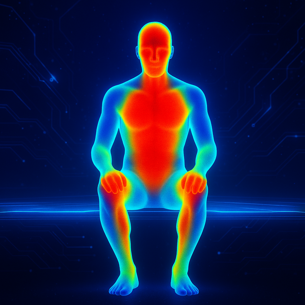
DITI isn’t just a diagnostic snapshot — it’s a dynamic tool that supports the entire lifespan of an athletic injury. From the very first sign of discomfort to the final clearance for competition, thermography plays an active role in helping clinicians make smart, data-driven decisions. Because it visualizes subtle physiological changes such as heat patterns, inflammation, vascular shifts, and sympathetic responses, DITI gives providers an objective window into what the athlete’s body is doing beneath the surface long before structural imaging reveals anything significant.
At the earliest stages of an injury, thermography can detect abnormal thermal patterns that suggest irritation, overuse, or emerging dysfunction. These physiologic “warning signals” allow clinicians to intervene early — often preventing a minor issue from progressing into a more serious strain, sprain, or chronic condition. As treatment begins, DITI becomes a valuable comparative tool, creating a visual record of improvement or stagnation that guides therapy choices and helps tailor rehabilitation.
During the recovery phase, athletes often feel pressure to return to play quickly, even if deeper physiological disruptions remain. Thermography helps remove the guesswork by providing objective evidence of whether inflammation has resolved, circulation has normalized, or autonomic responses have stabilized. This insight allows providers to confidently determine whether the injured area is genuinely healing or still at risk.
By the time return-to-play evaluations occur, DITI offers clinicians an additional layer of certainty. When paired with physical assessment and functional testing, thermography delivers a clearer, more complete picture of readiness — supporting safer decisions and reducing the likelihood of re-injury.
DITI doesn’t just record injuries; it actively informs every stage of care.
DITI plays multiple roles throughout recovery, including:
-
Confirming a diagnosis by identifying abnormal thermal patterns that correlate with inflammation, nerve irritation, or vascular changes.
-
Identifying injury patterns early, even before structural changes appear on X-rays or MRIs.
-
Tracking healing over time, offering a visual timeline of progress that both athletes and clinicians can clearly understand.
-
Assessing treatment effectiveness, allowing providers to adjust therapies based on the body’s real-time response.
-
Predicting return-to-play readiness with objective markers that reduce guesswork and help prevent re-injury.
Because DITI focuses on function rather than structure, it excels at detecting physiological reactions like changes in blood flow, sympathetic nervous system activation, and localized inflammation. This makes it especially useful for conditions involving soft tissue injury, nerve dysfunction, autonomic imbalance, or chronic pain — areas where traditional imaging often falls short.
Frequently Asked Questions
- Can thermal imaging (DITI) show early signs of overuse or ‘pre-injury’ in athletes before they feel pain?
- How does DITI compare or complement other technologies like ultrasound, MRI or motion-analysis in a sports-medicine clinic?
- Are there limitations or situations where DITI is not appropriate or sufficient in sports-medicine use?
Q1: Can thermal imaging (DITI) show early signs of overuse or ‘pre-injury’ in athletes before they feel pain?
Answer: Yes — because DITI visualizes changes in skin-surface temperature driven by microcirculation and sympathetic nervous system activity, it can potentially pick up areas under silent stress (e.g., increased heat from inflammation or cooling from vasospasm) before full-blown symptoms appear. This means an athlete, coach or clinician could use regular DITI snapshots to monitor for emerging hotspots or cooling patterns, intervene early (alter training, apply therapy) and reduce risk of a major injury. It’s important to stress it’s a monitoring adjunct—not a guarantee—and must be paired with clinical judgement and other assessments.
Q2: How does DITI compare or complement other technologies like ultrasound, MRI or motion-analysis in a sports-medicine clinic?
Answer: DITI brings a unique, non-invasive “physiologic” perspective (thermal/emission patterns) rather than purely structural imaging. For example:
-
Ultrasound or MRI show anatomy (tissue tear, fluid, bone changes).
-
Motion-analysis shows biomechanics (how someone moves).
-
DITI shows autonomic/vascular responses (how the tissue behaves thermally under load or injury).
Because of this distinct viewpoint, DITI can complement other modalities: you might use DITI for screening or tracking progress (e.g., reduced abnormal hot/cold zones over time), and reserve structural tests when DITI raises a flag. It helps with quick, risk-free checks and progress monitoring when MRI/CT would be overkill or not yet indicated.
Q3: Are there limitations or situations where DITI is not appropriate or sufficient in sports-medicine use?
Answer: Absolutely — while DITI is powerful, it’s not a stand-alone replacement for all injury diagnostics. Some key limitations include:
-
It does not directly show structural damage (e.g., you won’t “see” a torn ligament on DITI).
-
Its accuracy depends on consistent test conditions (room temperature, skin prep, absence of recent exercise, etc.).
-
Certain underlying conditions (e.g., heavy scar tissue, very deep injuries, atypical vascular anatomy) may degrade thermal signals.
-
Some insurance or regulatory frameworks may not recognise DITI as a sole diagnostic criterion.
Thus, DITI works best as part of a multi-modal strategy: screening + monitoring + clinical exam + selective structural imaging when needed.
